 MOHS micrographic surgery
 technique was first developed by Dr. Frederic MOHS and is a specialized, highly effective method for removing skin cancers. The current modern and revised MOHS surgery technique differs from other skin cancer surgeries in that it permits the immediate and complete microscopic examination of the removed cancerous tissue, so that all “roots” and extensions of the cancer can be eliminated. Due to the methodical manner in which tissue is removed and examined, MOHS surgery has been recognized as the skin cancer treatment with the highest reported cure rate. MOHS technique is most commonly used to remove the 2 more common forms of skin cancers,
Basal Cell Carcinoma and
Squamous Cell Carcinoma.

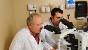
During the MOHS procedure, our doctors will remove the visible cancer with a narrow margin around it. The specimen is then processed in the MOHS lab in our office. Slides are subsequently prepared by our MOHS technician and reviewed by our doctors under the microscope. If the margins are cancer free, the wound can then be closed (small or superficial defects could sometimes be left open to heal on their own). However, if during the microscopic examination more cancer cells are discovered, extra tissue is removed from the positive margins. The process is repeated until no more cancer is seen.
Our doctors and their assistants will answer any additional questions you may have about this procedure.


